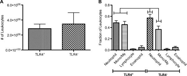
Fig. 4.
Adherent leukocyte profiles on PET discs following 16 h of IP implantation into TLR4+ or TLR4− mice. (A) Total number of adherent leukocytes collected from implants in either strain. No statistical difference was found between strains (p = 0.27); n = 7–9 mice per group. (B) Adherent leukocyte profiles showing fractions of neutrophils, monocyte/macrophages, eosinophils, and lymphocytes. Brackets indicate significant differences between groups (p < 0.05); n = 7–9 mice per group. All treatments significantly different from each other except for lymphocyte and eosinophil fractions from either strain as well as neutrophil and monocyte/macrophage fractions from TLR4+ strain.

