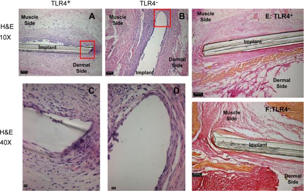
Fig. 6.
Representative H&E and Van Gieson stained tissue sections of PET discs implanted SC for 2 weeks. Shown are 10× images of representative H&E stained sections from a TLR4+ (A) or a TLR4− (B) mouse (bar indicates 100 μm) as well as 40μ magnifications TLR4+ (C) or a TLR4− (D) (bar indicates 10 μm). Also shown are 10μ images of Van Gieson (collagen) stained sections from a TLR4+ (E) or a TLR4− (F) mouse (bar indicates 100 μm). No noticeable differences in fibrous capsule formation were found between the two strains.

