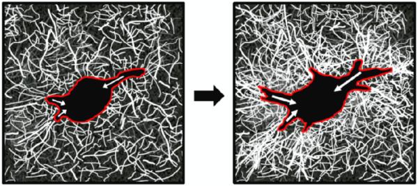
Fig. 5.
Cartoon schematic of ECM remodeling as a metric of force generation in 3D.
Cells (red outline) generate contractile forces (white arrows), which develop over time and contribute to local ECM remodeling, as indicated by increased collagen fiber density and orientation relative to the cell body. Collagen organization and density can by visualized using Confocal Reflectance Microscopy.

