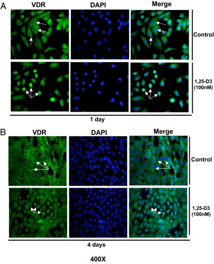
Fig. 2.
Expression and nuclear translocation of VDR upon incubation of C2C12 cells with 1,25-D3. Cultures of C2C12 cells were treated as in Fig. 1 in four-well removable chamber slides and subjected to IF using a polyclonal antibody for VDR followed by a FITC-conjugated secondary antibody (green). Cells were counterstained with DAPI (blue) to show nuclear localization. Merge pictures were done combining the green and blue pictures together to show nuclear translocation of VDR. Magnification, ×400. A, Cells incubated or not with 1,25-D3 for 1 d. B, Cells incubated or not with 1,25-D3 for 4 d.

