Fig. 1.
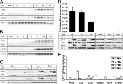
FGF21 initiates FGF signaling pathways in the liver in vivo. C57Bl6 mice were injected via the IVC with indicated quantities of FGF21. Tissue lysates were immunoblotted for total and phospho-FRS2 and ERK1/2. A, Robust increases in FRS2 and ERK1/2 phosphorylation in the liver in response to FGF21 at doses as low as 70 ng/g. B, Perigonadal WAT used as a positive control showing phosphorylation of FRS2 and ERK1/2 at similar doses to the liver. C, Time course. FGF21 (350 ng/g) was injected IVC and liver harvested at indicated time points. Blot shows increases in phosphorylation at time points as short as 5 min after IVC injection, with a maximal response occurring after 10 min. D, Immunoblots are shown of multiple tissues from mice injected IVC with either 350 ng/g FGF21 or saline. Depicted above is the mRNA expression of β-Klotho in these tissues as assessed by quantitative PCR. These data highlights the fact that FGF21 appears to activate FGF signaling only in tissues that express relatively high levels of β-Klotho. E, FGFR expression in peripheral tissues. Quantitative PCR data are shown normalized to 36B4 expression and given as mean ± sem.

