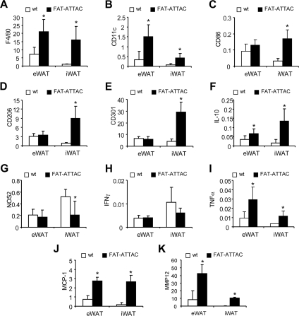
Fig. 2.
mRNA expression of macrophage markers is up-regulated upon apoptosis induction in FAT-ATTAC mice. Six-week-old wild-type (wt) (n = 5) or FAT-ATTAC (n = 6) mice were subjected to AP20187 dimerizer treatment for 14 d. Total RNA was isolated from epididymal (eWAT) and inguinal (iWAT) white adipose tissue. Quantitative PCR analysis was performed using primer pairs specific for macrophage markers (A) F4/80, (B) CD11c, (C) CD86, M2 macrophage markers (D) CD206, (E) CD301, (F) IL-10, M1 macrophage markers (G) NOS2, (H) IFNγ, (I) TNFα, and remodeling marker genes (J) MCP-1 and (K) MMP12. Data are represented as relative amount of mRNA expression normalized to hypoxanthine phosphoribosyltransferase. Each bar represents mean ± sd; *, P < 0.05 wild type vs. FAT-ATTAC.

