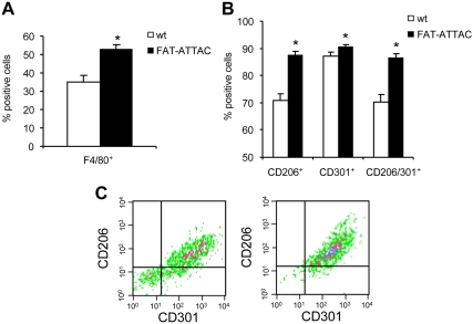
Fig. 4.
F4/80 macrophages recruited to adipose tissue upon fat cell apoptosis express the M2 markers, CD206 and CD301. Six-week-old wild-type (wt) or FAT-ATTAC mice were subjected to AP20187 dimerizer treatment for 14 d. SVC from epididymal adipose tissue were obtained by collagenase digestion. A, Quantification of F4/80 positive macrophages by flow cytometry. B and C, M2 macrophage marker expression. F4/80 macrophages were gated and analyzed for CD206, CD301, or CD206/CD301 expression. n = 4 mice per group. *, P < 0.05. C, Representative plot demonstrating CD206 and CD301 expression on F4/80 macrophages. Left, Wild-type; right, FAT-ATTAC.

