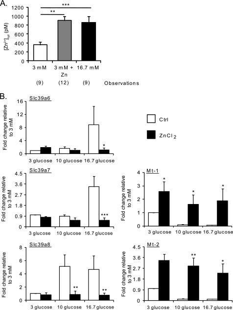
FIGURE 4.
Effect of extracellular Zn2+ on [Zn2+]cyt and on the expression of Slc39a6, Slc39a7, Slc39a8, Mt-1, and Mt-2. A, dissociated mouse islets were infected with eCALWY-4-expressing adenovirus and subsequently incubated for 24 h in medium containing 16.7 mm glucose or 3 mm glucose with or without 50 μm ZnCl2 before imaging on an Olympus IX microscope. [Zn2+]cyt was calculated as described under “Results” and in the legend to Fig. 1. Bars represent mean ± S.E. The number of cells imaged (n) on three different days of experiments is given in brackets under each bar. **, p < 0.01; ***, p < 0.001. B, mouse islets were incubated for 1 h at 3 mm glucose before culture at either 3, 10, or 16.7 mm glucose for 24 h in the presence (black bars) or absence (white bars) of 50 μm ZnCl2. qPCR analysis of Slc39a6, Slc39a7, Slc39a8, Mt-1, and Mt-2 was performed. Values of expression were normalized to that of a housekeeping gene (cyclophillin A). The fold changes compared with the values obtained at 3 mm glucose are plotted. Bars represent mean ± S.E. (n = 4). *, p < 0.05; **, p < 0.01; ***, p < 0.001.

