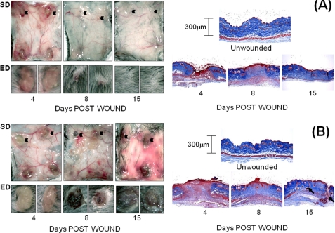
FIGURE 1.
Macroscopic view of dermal and epidermal wound closure in WT (A) and ts5−/− (B) mice. Four 3-mm wounds were made on the dorsal skin of mice that were sacrificed at 4, 8, and 15 days post-wounding. Left-hand panels show the macroscopic appearance of the dermal skin flap on the subdermal side (SD) to illustrate vascularization and inflammatory responses. The epidermal (ED) view of two representative wounds (indicated by black arrowhead) is also shown. Right-hand panels show representative histological sections after Mason trichrome staining of unwounded skin and wounds at 4, 8, and 15 days. Size bars for histology are given in the right-hand panels.

