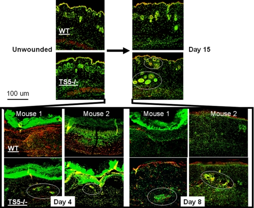
FIGURE 3.
Localization of aggrecan in unwounded skin and 4, 8, and 15 days post-wounding from WT and ts5−/− mice. Sections were stained for confocal microscopy as described under “Experimental Procedures.” Images shown were taken under ×10 magnification. Green fluorescence, nuclei; red fluorescence, aggrecan; yellow, intracellular or pericellular aggrecan. Aggrecan-+ve cell aggregates in ts5−/− wounds are shown by white circles. Also see supplemental Fig. S2 for nonimmune controls. Size bar, 100 μm.

