FIGURE 4.
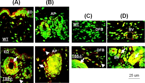
Localization of aggrecan in epidermal and dermal layers of ts5−/− and WT mice at 15 days post-wounding. Sections were stained as described under “Experimental Procedures” and viewed at ×40 to show the epidermal layer (ED) (A), newly formed skin appendages beneath the epidermal layer (B), dermal fibroblasts (DFB) (C), and subdermal blood vessels (BV) (D). AP, skin appendages. The accumulation of aggrecan in cell aggregates and fibroblasts in ts5−/− mice only is shown by white arrowheads. Green fluorescence, nuclei; red fluorescence, aggrecan; yellow, intracellular or pericellular aggrecan. Size bar, 25 μm size. Also see supplemental Fig. S3 for versican and hyaluronan localization in ts5−/− cell aggregates.

