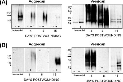
FIGURE 6.
Aggrecan and versican deposition in regenerating skin of Cd44−/−;ts5−/− (A) and Cd44−/− (B) mice. Proteoglycans were prepared for Western analysis with anti-aggrecan (αDLS) and with anti-versican V1 (Ab1033). Each lane was loaded with 20% of the proteoglycan-rich fraction isolated from the four wound sites combined from one mouse. Samples loaded without and with chondroitinase ABC digestion are marked − and +, respectively. Images separated by dividing lines were from different parts of the same gel.

