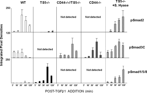
FIGURE 9.
Smad phosphorylation in newborn fibroblasts from WT, ts5−/−, ts5−/−/Cd44−/−, and Cd44−/− + S.Hyase mice. Fibroblast cultures from all genotypes were established and treated with TGFβ1 as described under “Experimental Procedures” (for basal matrix gene expression levels see supplemental Table 1). ts5−/− cells were also pretreated with Streptomyces hyaluronidase as described under “Experimental Procedures” before exposure to TGFβ1. Cell extracts were analyzed by Western for total and phosphorylated Smad2, Smad3C, and Smad1/5/8 (see typical gel images in supplemental Fig. S4). Data are expressed as integrated pixel density units for phosphorylated Smad proteins at 5–120 min post-TGFβ1 addition. Before the addition of TGFβ1, cells contained no detectable pSmad2 or pSmad 1/5/8. For two of the four WT cell preparations assayed, pSmad3C was detected even without addition of TGFβ1, and it represented about 25% that detected after 5 min. Data shown represent the mean ± S.D. for analyses of three separate cell preparations for each genotype.

