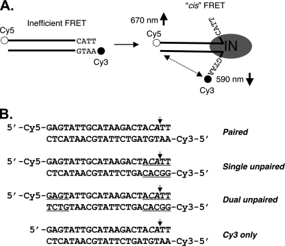
FIGURE 1.
DNA end fraying and experimental design. A, illustration of ASV viral DNA end fraying of terminal ∼4 bp by ASV IN as determined previously (4) and the predicted change in intramolecular (cis) FRET between donor (cyanine 3 (Cy3), filled circles) and acceptor (cyanine 5 (Cy5), open circles) fluorophores attached to the 5′ ends of a 22-bp oligodesoxyribonucleotide substrate. IN multimers, minimally dimers, are represented by a gray oval. B, sequences of the ASV-viral substrate duplexes used in these studies. Mismatched nucleotides at the termini are underlined. The small vertical arrows mark the normal sites of 3′ end processing by IN, in what is denoted the cleaved strand.

