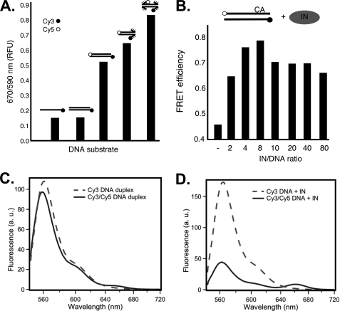
FIGURE 2.
FRET changes depend on disruption of base pairing in DNA termini and the ratio of ASV IN to substrate DNA. A, FRET measured in the absence of IN. The bars show relative FRET values obtained with single- or double-stranded ASV-DNA end duplex substrates that contain only a donor fluorophore and duplex oligonucleotides that contain both donor and acceptor fluorophores with paired, or singly or doubly unpaired ends. B, FRET increase of the paired ASV-DNA end duplex substrate, optimal in IN excess, peaking as the ratio approaches 4. Concentration of the paired ASV-DNA end oligonucleotide was kept constant at 50 nm. Sequence is shown in Fig. 1. C, fluorescence emission spectra of matching (50 nm) samples of ASV-DNA duplex labeled with donor only (Cy3, dashed line) or donor and acceptor (Cy3/Cy5, solid line). D, emission spectra of the same DNA samples as in C, but in the presence of 400 nm ASV IN. Instrumental parameters, including excitation wavelength (535 nm) and bandwidth (2 nm for excitation and 4 nm for emission), were identical in both C and D. Labeling conventions in A and B are as in Fig. 1, with CA above the top line in B representing the conserved dinucleotide in the cleaved strand.

