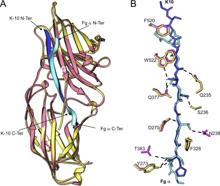
FIGURE 3.
Comparison of ClfB·K10 and ClfB·Fg α-chain peptide. A, ribbon representation of the superimposition of ClfB·K10 and ClfB·Fg α-chain complexes. ClfB from the K10 and Fg α-chain complexes is shown in yellow and pink, respectively. The K10 peptide is shown in blue, and the Fg α-chain is shown in cyan. B, comparison of the key interactions of Fg α-chain and K10 with ClfB. The ClfB molecules of the two complexes were superimposed and are not shown for clarity. K10 peptide is shown in blue and the Fg α-chain peptide is shown in cyan. The side chains of ClfB from the K10 complex are shown in yellow, and those from Fg α-chain complex are shown in pink.

