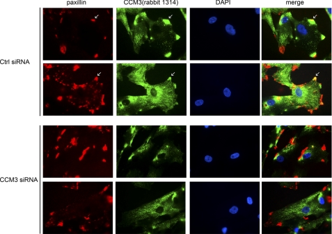
FIGURE 4.
Co-localization of endogenous CCM3 and paxillin in pericytes. Pericytes were transfected with control (Ctrl) or CCM3 siRNA. 48 h post-transfection, cells were immunostained with anti-CCM3 and paxillin followed by counterstaining with DAPI (blue) for nuclei. Merged images are shown on the right. Representative images from 10 cells in each group are shown. Scale bar, 20 μm.

