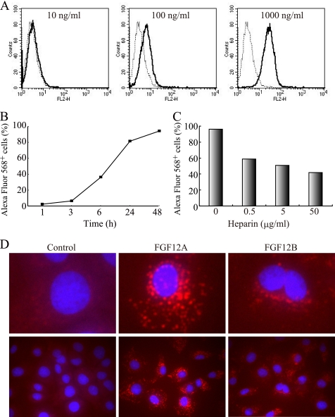
FIGURE 2.
Internalization of FGF12 into IEC6 cells. Human recombinant FGF12 was labeled with Alexa Fluor 568. FGF12B (R&D) had no tags, whereas FGF12A was linked to a 3xFLAG-His6 tag as described previously (32). A rat intestinal epithelial cell line, IEC6, was cultured in complete medium with Alexa Fluor 568-labeled FGF12 A, IEC6 cells were incubated with FGF12B at a dose of 10, 100, or 1000 ng/ml for 24 h. They were harvested in trypsin-EDTA, washed twice with phosphate-buffered saline (PBS) containing 0.2% bovine serum albumin (BSA), and subjected to FACS Calibur flow cytometry to estimate fluorescence intensity. A dotted line shows the control cells and a solid line shows the FGF12-treated cells. B, kinetics of cellular uptake of extracellular FGF12 was examined over 48 h by flow cytometry. IEC6 cells were incubated in culture medium with 1 μg/ml of FGF12B and subjected to flow cytometry to determine the percentage of Alexa Fluor 568-positive cells at the indicated time points. C, IEC6 cells were cultured for 24 h in medium with 1 μg/ml of FGF12B and 0.5, 5, or 50 μg/ml of heparin. They were subjected to flow cytometry. D, IEC6 cells were cultured for 24 h in medium with 1 μg/ml of FGF12A or FGF12B. The cells were fixed in 1% glutaraldehyde, and the nuclei were visualized by staining with 20 μg/ml of Hoechst 33342 (blue) and the fluorescence confocal images were acquired using a IX81 fluorescence microscope with a disk scanning unit (Olympus, Tokyo, Japan).

