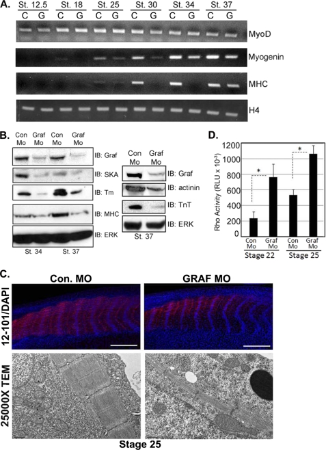
FIGURE 12.
GRAF1 morphants exhibit elevated RhoA activity and impaired skeletal muscle differentiation. A, RNA from Con Mo (C)- and GRAF1 Mo (G)-injected embryos (n = 10) was isolated and utilized for semi-quantitative RT-PCR analysis of indicated marker gene. B, lysates from Con Mo- and GRAF1 Mo-injected embryos at the indicated stages were analyzed by Western analysis. Note that the lysates shown in the left panel are identical to those shown in Fig. 2A. Lysates used for right panel were collected from a separate experiment. IB, immunoblot. C, top, laser scanning confocal microscopy of whole-mount 12–101 (red) and ToPro3 (blue)-stained stage 25 Con Mo- and GRAF1 Mo-injected embryos (scale bar, 500 μm). Note appropriate alignment of nuclei but reduced skeletal muscle differentiation (also see Fig. S3, A and B and Table 1). Bottom, TEM (×2500 magnification) from somite-matched stage 25 Con Mo- and GRAF1 Mo-injected embryo. Note reduced myofiber content in GRAF1 morphants relative to controls. D, ELISA-based RhoA activity assays were performed on lysates isolated from stage 22 and 25 Con Mo- and GRAF1 Mo-injected embryos. Ten embryos were processed in batch for each stage and treatment.

