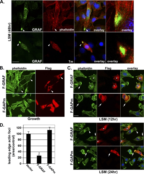
FIGURE 7.
GRAF1 is recruited to a pre-fusion complex and promotes actin-foci dissolution. A, C2C12 cells were exposed to differentiation conditions (LSM) for 48 h, and endogenous GRAF1 was detected by confocal immunofluorescence microscopy. Note high level of GRAF1 protein localized to the tips of pre-fused myoblasts. Co-staining with phalloidin to detect filamentous actin or Tm reveals lack of actin-based structures in the GRAF localization domain (arrowheads). Scale bar, 50 μm. B and C, L6 myoblasts transfected with F-GRAF or F-GAPm in growth media or LSM for the indicated time points. Cells were co-stained with anti-FLAG antibody and phalloidin. Overlay shows high magnification merge of GRAF1 variants and phalloidin. Scale bars, 50 μm. D, quantification of areas of actin foci at the leading edge of F-GRAF and F-GAPm cells is shown graphically (n = at least 100 cells/condition from 3 experiments). *, p <0.05.

