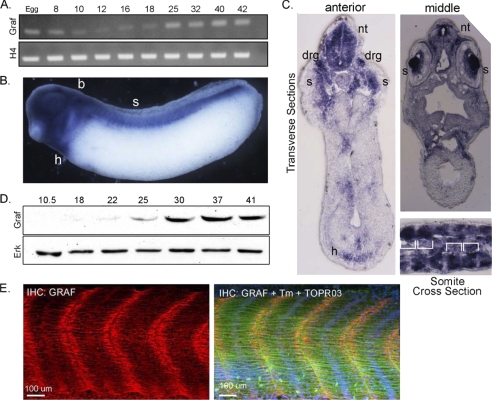
FIGURE 9.
GRAF1 is highly expressed during somitogenesis. A, RT-PCR analysis for x.GRAF1 and histone H4 (H4; loading control) was performed using RNA isolated from embryos at the indicated stages. B and C, whole-mount in situ hybridization of stage 29 Xenopus embryo using an antisense probe specific for x.GRAF1: h, heart; b, brain; s, somites; nt, neural tube; drg, dorsal root ganglia. B, lateral view. C, top, transverse; bottom; cross-section through mid-somite region (somite borders indicated with brackets). D, Western blot analysis of GRAF1 and ERK (loading control) expression at the indicated developmental stages. E, laser scanning confocal images of whole-mount GRAF1 (red), tropomyosin (green), and ToPro3 (blue nuclei) stained somites from wild type stage 37 embryo. IHC, immunohistochemistry.

