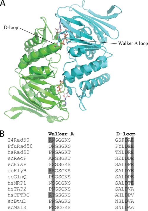
FIGURE 1.
A, ribbon structure of the dimeric PfuRad50 nucleotide binding domain. The subunits of the dimer (PDB code 1F2U) are colored green and cyan with the ATP molecules located at the dimer interface. The Walker A and D-loop motifs from opposing subunits that form a single ATP active site are indicated. B, multiple amino acid sequence alignment of the Walker A and D-loop motifs from selected Rad50 and ABC proteins. The protein names are preceded by the organism abbreviations, which are as follows: T4, bacteriophage T4; Pfu, P. furiosus; hs, Homo sapiens; and ec, E. coli. The dark gray shading indicates the conservation of the residues mutated in this study.

