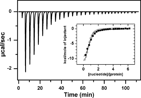
FIGURE 1.
Half of the nucleotide binding sites of WT ClpB bind ATPγS or ADP below 50 μm nucleotide. Shown is the base-line-corrected instrumental response of successive injections of ATPγS into a solution of WT ClpB at 25 °C. 6 μl (1.6 mm nucleotide) were injected into a solution containing 28 μm WT ClpB. Experiments were performed in 50 mm Tris-HCl, pH 7.5, 150 mm KCl, 20 mm MgCl2. Inset, shown are the integrated data of WT ClpB titration with ATPγS (black squares) or ADP (white circles) and fit of the corresponding binding isotherms to a single-site binding model (solid lines).

