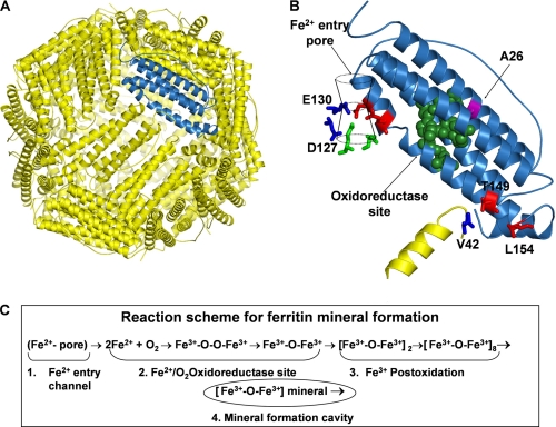
FIGURE 1.
Ferritin nanocage structure. A, frog-M ferritin nanocage structure, viewed from the outside with the 3-fold axis near the center, shown with one subunit highlighted in blue (Protein Data Bank ID code 1MFR). Eight Fe2+ ion channels (10), each formed by residues from three subunits around the 3-fold axes, lead through the protein cage from the outside of the cage to the large (8-nm diameter) central cavity, near several residues of the active sites; B, a single subunit of the frog-M ferritin showing all of the residues studied by site-directed mutagenesis; C, steps in ferritin mineral synthesis.

