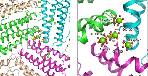
FIGURE 3.
Distribution of metal ion in ferritin protein cages to three active sites depends on the structure of the ion channels at the 3-fold cage axes. Three Mg2+ ions (hexahydrated) are observed bound to the Asp127 carboxylate and Glu130 carbonyl from each of the three frog-M ferritin near the ion channel exits into the ferritin cavity; the view is from inside the cavity into the ion channel toward the external pore opening in the exterior cage surface. In addition, in the center of the ion channel are three of the four hexahydrated Mg2+ in the line that stretches from the outside of the cage to the cavity entrance (10). The figure was made from Protein Data Bank ID code 3KA3; Mg, green spheres; coordinated water, red spheres; fragment of backbone of helix 3 of subunit forming the iron channel subunit 1, green; subunit 2, magenta; subunit 3, azure.

