
Figure 1.
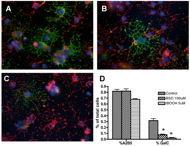
Oligodendrocyte differentiation after 72 hours of oxidant exposure. OPCs were double-labeled in vitro with oligodendroglial markers A2B5 (red) and GalC (green) after 72 hours of oxidant exposure. The number of mature GalC+ cells was significantly decreased in the BSO (B) and tBOOH (C) exposed cultures compared to control (A) cultures. The majority of cells exposed to oxidative stress were arrested in the immature A2B5+ cell stage. Quantitative analysis of A2B5+ and GalC+ cells is shown in (D). The proportion of positive cells per 40× field was determined by counting 10 fields per coverslip from three separate experiments following indirect immunofluorescence using antibody to A2B5 and GalC. Data represent the mean plus standard deviation. *P < 0.05 vs. control.