
Figure 5.
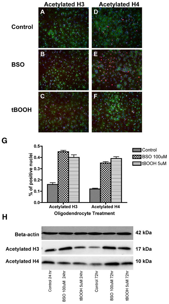
OPCs were stained in vitro with antibodies to acetylated histone H3 (A-C) and acetylated histone H4 (D-F) after 72 hours of exposure to oxidative agents. The density of acetylated histone H3 and H4 was significantly higher in BSO- and tBOOH-exposed cells. Quantitative analysis of acetylated histone H3+ and H4+ nuclei is also shown (G). The proportion of positive cells per 20× field was determined by counting 10 fields per coverslip in three separate experiments following indirect immunofluorescence using antibody to acetylated histone H3 or H4. Data represent the mean plus standard deviation. *P <0.05. Western blotting was performed on OPC lysate after 24 and 72 hours (H). Analysis reveals an increase in acetylated histone H3 and H4 protein after 72 hours of exposure to oxidative stress. Each lane contains 25 μg protein.