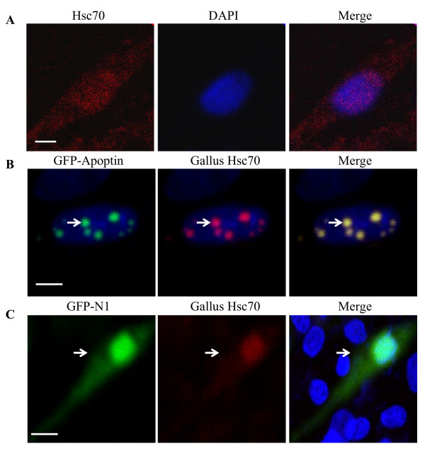
Figure 6.
Localization of Apoptin and endogenous Hsc70. (A) Localization of endogenous Hsc70 in DF-1 cells. DF-1 cells were seeded on 24-well plates with coverslips and cultured overnight. The cells were fixed with 1% paraformaldehyde. After washing, the fixed cells were permeabilized with 0.1% Triton X-100, and probed with anti-Hsc70 and TRIC-conjugated secondary antibodies. Nuclei were counterstained with DAPI (Blue). (B) Colocalization of endogenous Hsc70 with Apoptin in the nucleus. DF-1 cells were transfected with a pEGFP-Apoptin plasmid for 24 hours and immunostained as described above. (C) Negative control pEGFP-N1 vector was transfected into DF-1 cells for 24 hours and immunostained as described above. The cell samples were observed with a laser confocal scanning microscope. The scale bars represent 10 μm.

