Figure 5.
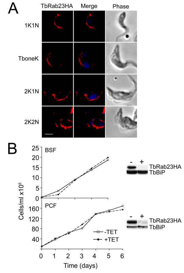
TbRab23 location is conserved in the PCF and expression is nonessential for proliferation of BSF and PCF cells. (A) A PCF strain constitutively expressing HATbRab23 fused to HA was stained with anti-HA and counterstained with Alexa-568 (red) to indirectly visualise TbRab23HA, together with DAPI to image the nucleus and kinetoplast. The location of TbRab23 in PCF was followed through the stages of the cell cycle, shown to the left of the images. Scale bar 2 μm. (B) Cumulative proliferation curves of BSF TbRab23RNAi cells (upper panel) and PCF TbRab23RNAi cells (lower panel) grown in the presence (broken line) or absence (solid line) of 1 μg/ml tetracycline. Silencing was validated at 24 hours post-induction by Western blot on RNAi cells expressing recombinant TbRab23, shown to the right of each graph. TbBiP was used to ensure accurate loading and specificity. "-" denotes uninduced and "+" induced cells.

