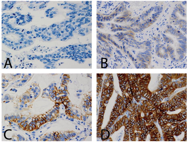
Figure 3.
HER2 immunohistochemical staining showing 0 immunostaining (A, ×400), 1+ immunostaining (B, × 400), 2+ immunostaining (C, × 400) and 3+ immunostaining (D, × 400) in esophageal adenocarcinoma. Both 2+ and 3+ uniform staining are considered as HER2 protein overexpression. Incomplete membranous “U” shape stain is presented in B.

