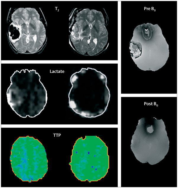
Figure 3. Advanced MRI of lobar intracerebral haemorrhage.
Left: before craniotomy. Middle: after craniotomy for treatment of mass-effect and removal of haematoma. Sequential T2, lactate magnetic resonance spectroscopy, and perfusion studies showed qualitative decreases of perihaematomal oedema and perihaematomal lactate and increased occipital regional perfusion measured as time to peak of bolus injectate (TTP) after removal of clot; TTP is represented by intensity and distribution of green colour. Right: magnetic susceptibility images show paramagnetic influence before surgery and limited susceptibility after removal of the iron-containing blood clot by craniotomy. Figures provided by J Ricardo Carhuapoma (Johns Hopkins Medical Institution, Baltimore, MD, USA).

