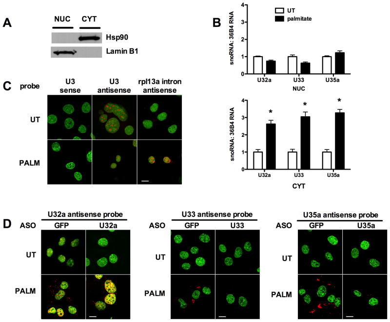
Figure 6. U32a, U33 and U35a accumulate in the cytoplasm under lipotoxic conditions.
(A, B) C2C12 cells were untreated (UT) or treated with palmitate for 24 h. Cells were separated into cytosolic (CYT) and nuclear (NUC) fractions by sequential detergent solubilization. (A) Fractions were analyzed by western blotting for a cytosolic marker, hsp90, and for a nuclear marker, lamin B1. (B) Total RNA was prepared from the fractions and analyzed for rpL13a snoRNA abundance relative to 36B4 by qRT-PCR. Graphs show mean ± SE from a representative experiment (n = 3). * p < 0.05 for palmitate treated vs. UT. (C, D) C2C12 cells were analyzed by in situ hybridization under basal conditions (UT) and following 24 h treatment with palmitate (PALM) using specific snoRNA or control probes (red). Nuclei were stained with SYTOX Green. (C) Cells were probed with U3 antisense probe for known nucleolar snoRNA, control U3 sense probe, and control rpL13a intron 1 antisense probe. (D) Control GFP ASO-nucleofected and specific snoRNA ASO-nucleofected cells were examined by in situ hybridization with antisense probes for U32a, U33 and U35a. Bars, 10 μm. (See also supplemental Figure 6.)

