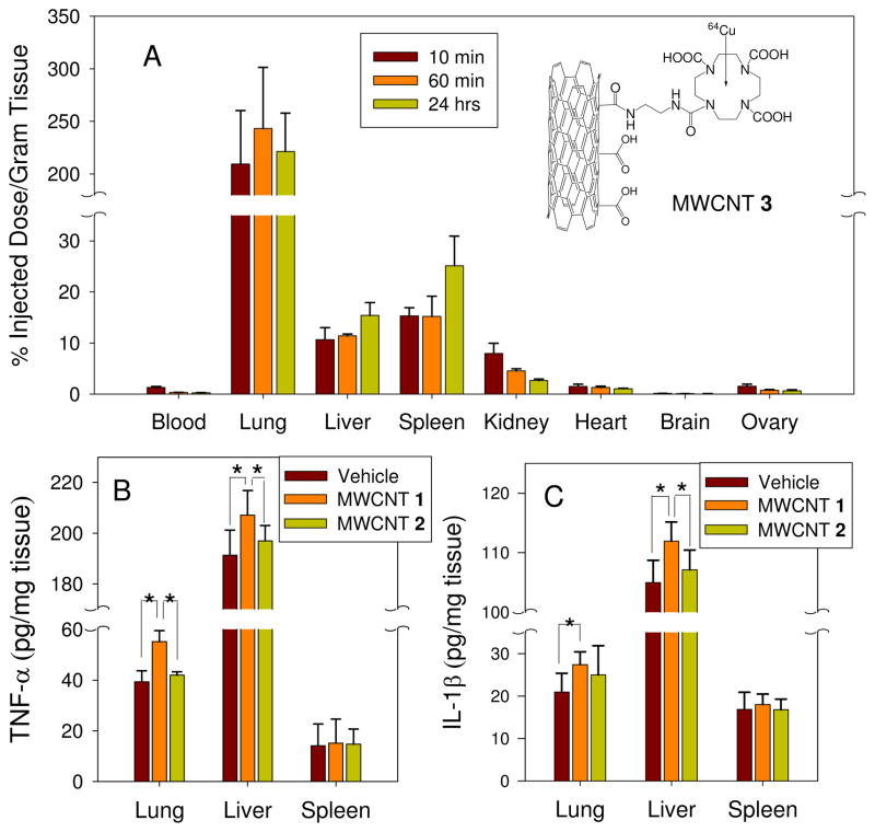
Figure 2.
The distribution of MWCNTs in mice and the effect of MWCNT 1 and 2 on inflammatory cytokines in mice lung, liver and spleen in vivo. (A): MWCNT 1 were labeled with a specific ligand for 64Cu. Suspension of MWCNT 1 was injected in to CD-1 female mice (4 mice per group) via tail vein. For 10 min, 60 min and 24 hrs, the accumulation of MWCNT 1 in the tissues were measured. (B and C): Suspension of MWCNT 1 and 2 in PBS solution in the presence of 0.1% Tween 80 was injected in to female BALB/c mice (5 mice per group) via tail vein with a dose of 14 mg/kg. For 24 hrs, TNF-α and IL-1β in the tissue homogenate were determined by ELISA. (*p<0.05)

