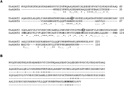
FIGURE 4.
Alignment of SaADATa and TbADAT2 suggests differences in tRNA binding. (A) Sequence comparison between the adenosine deaminases acting on tRNA of S. aureus and T. brucei shows that a number of key residues involved in substrate binding by the bacterial enzyme have naturally undergone nonconservative changes in the T. brucei enzyme. The amino acid sequences of T. brucei ADAT2 and S. aureus ADATa were aligned using Clustal W. Conservative changes are denoted by dots [(.) and (:)] placed under the sequences and identical residues by an asterisk (*). Amino acids involved in RNA interaction in the cocrystal of SaADATa and an anticodon stem–loop (ASL) representing the anticodon arm of S. aureus tRNAArg are shown in boldface letters. The amino acids in TbADAT2 shown within boxes have been mutated to alanine and are shown to play no role in tRNA binding in T. brucei. (B) The amino acid sequence of TbADAT2 was also analyzed for potential RNA binding domains by the RNAbindR program, which uses a naive Bayesian classifier for all predictions, as described by Terribilini (Terribilini et al. 2007). Potential domains are denoted by plus (+) signs under the sequence, and boldface letters denote the KR-domain which has been analyzed further in this study.

