Fig. 1.
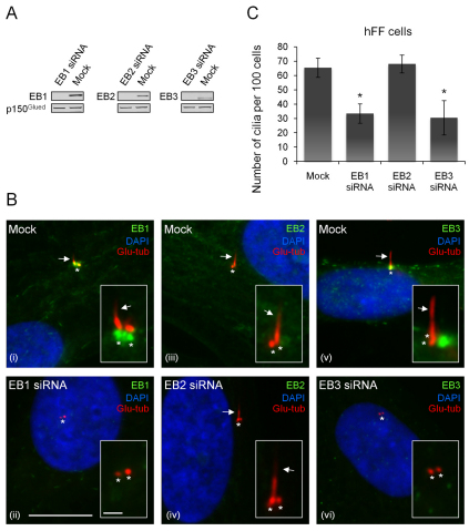
Depletion of EB1 or EB3, but not EB2, inhibits cilia formation. Human foreskin fibroblasts (hFFs) were treated with mock or EB-specific siRNA, as indicated, grown to confluency and serum starved for 48 hours. (A) Western blot analysis of lysates of mock or EB-siRNA-treated cells. Dynactin subunit p150Glued serves as loading control. (B) IFM images of methanol-fixed cells stained with Glu-tub antibody (red) and with rat monoclonal EB1, EB2 or EB3 antibodies (green) (Komarova et al., 2005). DNA was stained with DAPI (blue). Arrows indicate primary cilia, asterisks mark centrioles. Insets are enlarged images of the cilium or centriole region (shifted overlays). Scale bars: main panels, 10 μm; insets, 3 μm. (C) Quantitative analysis of IFM results. Each column represents the average of three independent experiments with at least 100 cells counted per sample each time; error bars represent standard deviation (s.d.). *, significantly different from mock control (P<0.01). Similar results were obtained in RPE cells (percent ciliated cells were: mock, 66±1%; EB1 siRNA, 30±1%; EB2 siRNA, 66±1%; EB3 siRNA, 34±10%).

