Fig. 6.
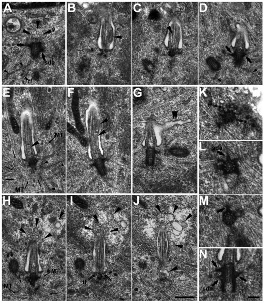
Ultrastructural defects in cells lacking EB3. TEM analysis of growth-arrested, EB3-depleted hFF cells. (A) Longitudinal section of a mother centriole with subdistal appendages (sda; thick arrow). At the distal end of the centriole three vesicles are visible (v; large arrowheads). Rootlet filaments (rf) are labeled (small arrowheads). (B–D) Consecutive serial longitudinal sections of a basal body with a stumpy cilium. Vesicles (large arrowheads) are visible within the cilium. In D, the oblique sectioned daughter centriole, subdistal appendages (thick arrows) attached to the basal body and rootlet filaments (small arrowheads) can be seen. (E,F) Consecutive serial longitudinal sections of a basal body with stumpy cilium. Vesicles (large arrowheads) are accumulated within the cilium. (G) Longitudinal section of a basal body bearing a stumpy cilium. The cilium pit shows a long, tubular extension (large double arrowheads). The oblique sectioned daughter centriole is visible to the right. (H–J) Consecutive longitudinal serial sections of a basal body with stumpy cilium. The cytoplasmic area around the tip of the cilium looks distorted and many vesicles (large arrowheads) are present. In J, vesicles (large arrowhead at the bottom of the panel) can be seen, which are localized in the region of the attachment site between the transition fibers (tf; thin double arrows in I) and the membrane of the cilium pit. (K–M) Consecutive serial cross sections of a mother centriole. The transition fibers (tf; thin double arrows) attached to the distal end of the centriole are clearly visible (K). At least six subdistal appendages are associated with the centriole in a subdistal position (L, sda; thick arrow); one additional subdistal appendage is associated with the centriole in a more median to proximal position (M, thick arrow). (N) Longitudinal section of a basal body. At the left side of the centriole two subdistal appendages (sda; thick arrows) are associated, above each other, with the centriole; at the right side only one subdistal appendage (thick arrow) is present. Scale bars: J, 500 nm (applies to A-J); N, 200 nm (applies to K–N).

