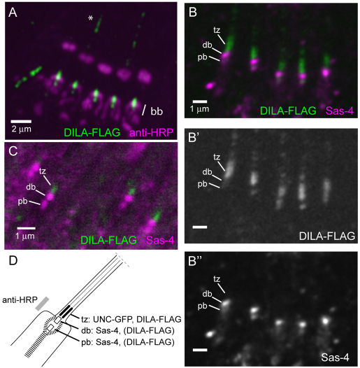
Fig. 5.
DILA–FLAG resides at the base of the cilium. (A) When expressed in larval Ch neurons, DILA–FLAG is localised at the basal body (bb) region, as indicated by anti-HRP antibody staining. The DILA–FLAG staining in the distal cilia (*) varies with the expression level and is presumed to be an artifact of protein overexpression. (B–B″) Sas-4 (magenta) reveals the distal and proximal basal bodies (db, pb) at the base of each cilium. DILA–FLAG staining overlaps with Sas-4 staining and is especially prominent in the cilium beyond the distal basal body, predicted to be the putative transition zone (tz). (C) When expressed at a low level, DILA–FLAG is confined to the putative transition zone. (D) Schematic of the cilium base showing the distribution of proteins.

