Fig. 7.
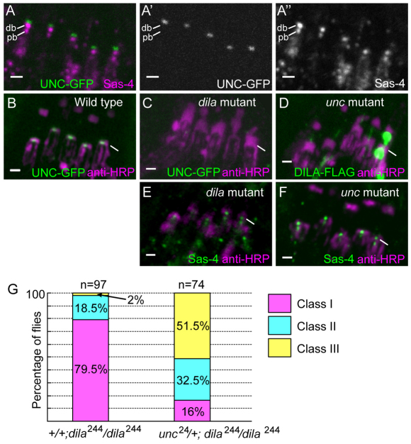
Co-dependent localisation of DILA–FLAG and UNC–GFP. (A–F) Larval Ch neurons. (A) The UNC–GFP fusion protein (green) is detected in the region just above the distal basal body, as marked by Sas-4 expression (magenta). This is predicted to be the transition zone. (B) UNC–GFP in wild-type flies, co-stained with anti-HRP (magenta). (C) In dila81 mutants, UNC–GFP is absent. (D) In unc24 mutants, DILA–FLAG staining is weak and diffuse, with little detectable in the basal body region (compare with Fig. 5B), except for some abnormal aggregations in one dendrite. By contrast, Sas-4 is still localised in dila (E) and unc (F) mutants. (G) The uncoordinated phenotype of the weak hypomorph dila244 is enhanced after mutation of one copy of unc. The phenotype was scored according to three classes of increasing severity of uncoordination (see Materials and Methods). The difference in distributions is significant according to a χ-squared test, P<0.001.

