Fig. 7.
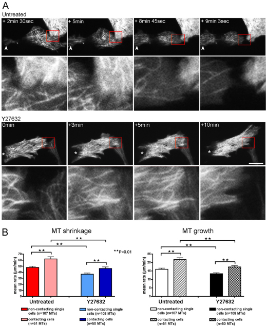
Microtubule dynamics changes at the leading edge of cells during cell–cell collision. (A) Top panels show a YFP–tubulin-expressing untreated CEF colliding with the lamella of the cell above and rapidly switching polarity (supplementary material Movie 9). Higher magnifications of the region of cell–cell contact (red box) are shown below their corresponding images. MT filaments flood into the tail end of the cell (white arrowhead) and away from the site of cell–cell contact to form a new leading edge, and subsequently, the cell is able renew migration in the opposite direction. Bottom panels show a YFP–tubulin-expressing Y27632-treated CEF colliding with the lamella of the cell below right (supplementary material Movie 10). The cell does not recoil away, and MTs are not lost from the region of cell–cell contact. Higher magnifications of the region of cell–cell contact (red box) are shown below their corresponding images. Asterisk denotes the tail end of the cell that is maintained throughout the collision episode. Scale bar: 18 μm. (B) Mean rates of MT shrinkage and growth at the region of cell–cell contact in untreated and Y27632-treated CEFs (from four different untreated CEFs, n=51 shrinking and n=53 growing filaments; three different Y27632 CEFs, n=50 shrinking and n=51 growing filaments; **P<0.01).

