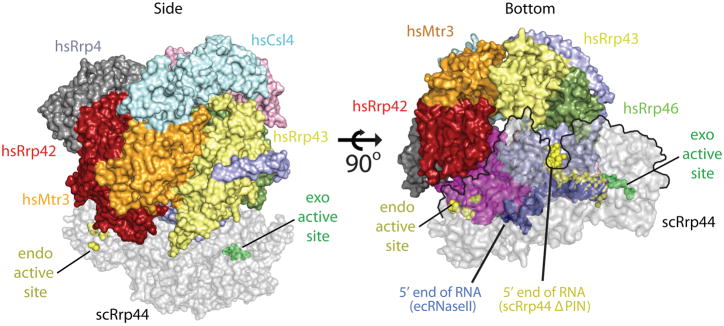
Figure 6. Model for RNA recruitment to the hydrolytic active site of Rrp44 within the eukaryotic exosome.
A 10-component exosome model was created by aligning the S. cerevisiae Rrp41-Rrp45-Rrp44 trimer (PDB ID = 2WP8) onto the Rrp41-Rrp45 subunits of the human exosome (PDB ID = 2NN6). Coloring for the 9-component exosome is described in Fig. 2, and the Rrp44 component is shaded grey. The left panel depicts a side view of the complex with the Rrp44 exoribonucleolytic active site indicated as green spheres, and the endoribonucleolytic active site as yellow spheres. The right panel depicts a bottom view of the complex. The RNA complexes determined for RNase II (blue spheres, PDB ID = 2IX1) and Rrp44ΔPIN (yellow spheres, PDB ID = 2VNU) were superimposed into the full-length Rrp44 structure to illustrate the paths of RNA in the complex. The Rrp44 molecule is outlined by a black line in the right panel where it overlaps with the exosome core.

