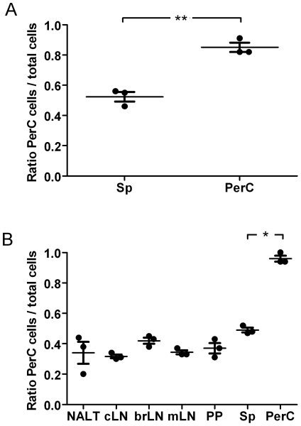
Figure 5. Peritoneal CD4+ T cells preferentially re-enter the peritoneal cavity.
(A) CXCR3+ or (B) CXCR3− CD4+ T cells isolated from spleen or peritoneal cavity were differentially labeled with CFSE or CMTMR, and mixtures containing equal numbers of cells were injected intravenously into naïve recipients. 18 hours after transfer, the ratio of transferred peritoneal cells to total transferred cells (peritoneal cells+spleen cells) was determined in different organs by flow cytometry. Similar results were obtained in three independent experiments. Dots represent the ratio for individual animals. Horizontal lines indicate the mean for the group, and error bars correspond to the SEM. The differences were statistically significant at P<0.0001 (*) and P<0.002 (**). NALT, nasal associated lymphoid tissues; cLN, cervical lymph node; brLN, bronchial lymph node; mLN, mesenteric lymph node; PP, Peyer's patches; Sp, spleen; PerC, peritoneal cavity.

