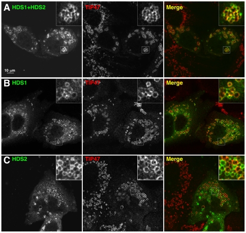
Figure 8. The GBF1 HDS1 and HDS2 domains localize to lipid droplets in cells.
HeLa cells transfected with Venus-tagged HDS1 and/or HDS2 domains of GBF1 as indicated were treated with 400 µM oleic acid for 3 hours, then immunostained with antibodies against TIP47 (middle panels). Venus fluorescence is shown on the left; the merge image on the right. Bar: 10 µm. A- Cells expressing the region of GBF1 from the beginning of HDS1 to the end of HDS2, N-terminally tagged with Venus. B- Cells expressing the Venus-tagged HDS1 domain. C- Cells expressing Venus-HDS2. Bar, 10 µm.

