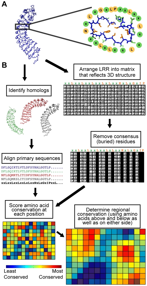
Figure 1. LRR structure and an outline of conservation mapping procedure.
(A) Left: a representative LRR domain (left), P. vulgaris PGIP2, which forms the regular spiral pattern typical of LRRs. Right: a single 24 amino acid repeat of the LRR, surrounded by circles designating the residues of the LRR consensus amino acid sequence (xxLxxLxxLxxLxLxxNxLxGxIP). Note that the consensus residue side chains (orange) form the core of the protein, whereas the variable residues (green) are solvent-exposed. (B) Schematic representation of the conservation mapping procedure, using the example of PGIP1-4. See Methods and Text S1 for a detailed description of the procedure.

