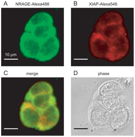
Figure 1. Endogenous NRAGE and XIAP expression in P19 cells.
P19 cells were fixed with 4% PFA and permeabilized followed by application of primary antibodies for NRAGE and XIAP and widefield imaging. Secondary Alexa 488 and 546 antibodies were used to identify NRAGE (A) and XIAP (B) respectively. (C) Merged imaging shows NRAGE and XIAP mainly occupy the cytoplasm with smaller concentrations in the nucleus. (D) Phase contrast image.

