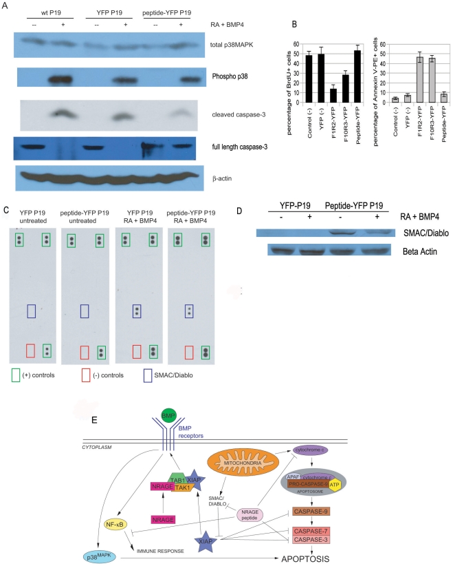
Figure 8. Apoptosis is inhibited in P19 cells cultured with the NRAGE peptide.
(A) Western blot of P19 cells transfected with the NRAGE peptide-YFP had less detection of cleaved and more full-length caspase-3 compared to wild-type or YFP transfected cells after 24 hours exposure of 1 µm RA and 10 ng/ml BMP-4. c. Higher levels of total-p38MAPK are detected in all cells that received apoptosis treatment. β-actin was used for loading control. (B) Stably integrated YFP and NRAGE peptide-YFP cells without retinoic acid (RA) and BMP-4 were lysed and incubated to human apoptosis profiler antibody nitrocellulose arrays (left 2 panels). Arrays were stripped and semi-stable YFP and NRAGE peptide-YFP cells incubated with 1 µM RA+10 ng/ml BMP-4 for 24 hours were lysed and incubated to the same respective arrays and found to bind with SMAC/Diablo, but with less detection for NRAGE peptide-YFP cells. (C) Western blot analysis of total cell lysate of SMAC/Diablo expression of YFP and NRAGE peptide-YFP cells with and without BMP-4 stimulation. (D) Annexin V and BRDU flow analysis of YFP control cells, NRAGE full length, NRAGE repeat, and NRAGE-peptide cell lines (E) Proposed signaling BMP MAPK pathway incorporating the inhibition of SMAC/Diablo by the NRAGE peptide resulting in the inhibition of cleaved caspase-3 and apoptosis.

