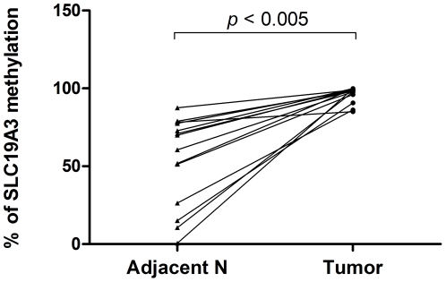
Figure 2. Increased percentage of SLC19A3 DNA methylation in primary breast cancer tissues.
Percentage of SLC19A3 promoter methylation between tumor tissues and their paired adjacent non-tumor breast tissues from the 15 breast cancer patients by MS-qPCR. Percentage of methylation in tissue samples was calculated by the following equation: % meth = 100/[1+2ΔCt(meth-unmeth)]%. ΔCt(meth-unmeth) was calculated by subtracting the Ct values of methylated SLC19A3 signal from the Ct values of umnethylated SLC19A3 signal. Statistical difference was analyzed by Wilcoxon test, P<0.005.

