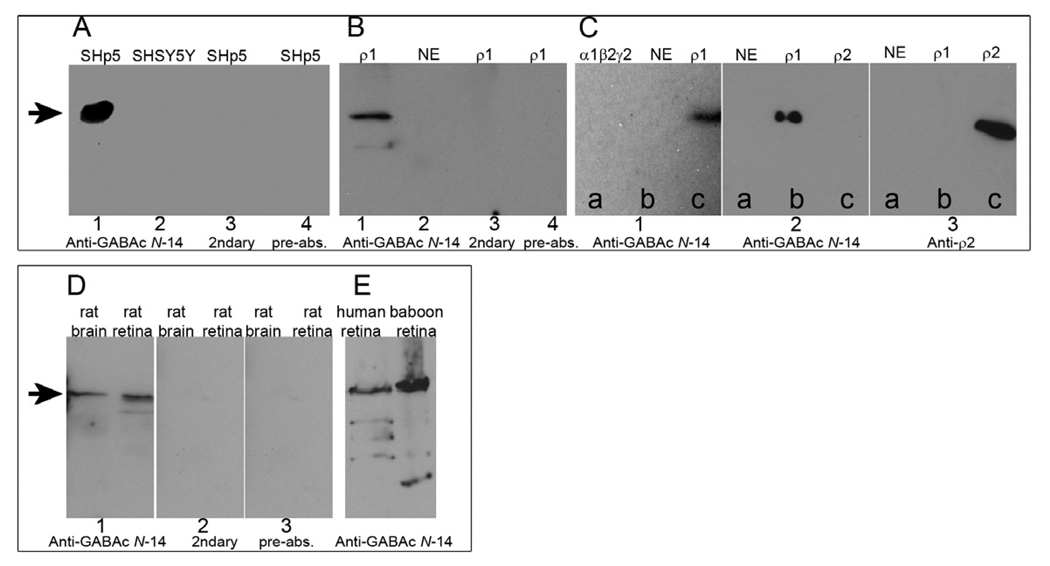
Fig. 2.
Western blots. A: Whole-cell lysates of GABAC-expressing SHp5-ρ1 cells and non-GABAC expressing SHSY5Y cells. Lane 1: SHp5-ρ1 probed with GABAC Ab N-14. Lane 2: SHSY5Y probed with GABAC Ab N-14. Lane 3: SHp5-ρ1 probed with secondary antibody only. Lane 4: SHp5-ρ1 probed with GABAC Ab N-14 pre-absorbed with N-14. B: Oocyte membrane preparations. Lanes 1, 3 and 4: GABAC ρ1 expressing oocytes. Lane 2: Non-expressing control oocyte. Experimental conditions used for lanes 1–4 are otherwise identical, respectively, to lanes 1–4 in A. C: Oocyte membrane preparations. Section 1: Preparations of α1β2γ2 GABAA-expressing oocytes (lane a), non-expressing oocytes (b), and ρ1 GABAC-expressing oocytes (c) probed with GABAC Ab N-14. Section 2: Preparations of non-expressing oocytes (lane a), ρ1 GABAC-expressing oocytes (b) and human ρ2 GABAC-expressing oocytes (c) probed with GABAC Ab N-14. Section 3: Same preparations as those of section 2, probed with anti-ρ2 antibody. D: Whole cell lysates of rat brain and rat retina probed with: GABAC Ab N-14 (Section 1), secondary antibody only (Section 2), or GABAC Ab N-14 pre-absorbed with N-14 (Section 3). E: Whole cell lysates of human and baboon retina, probed with GABAC Ab N-14. On all blots, the arrows indicate MW ~ 55 kDa.

