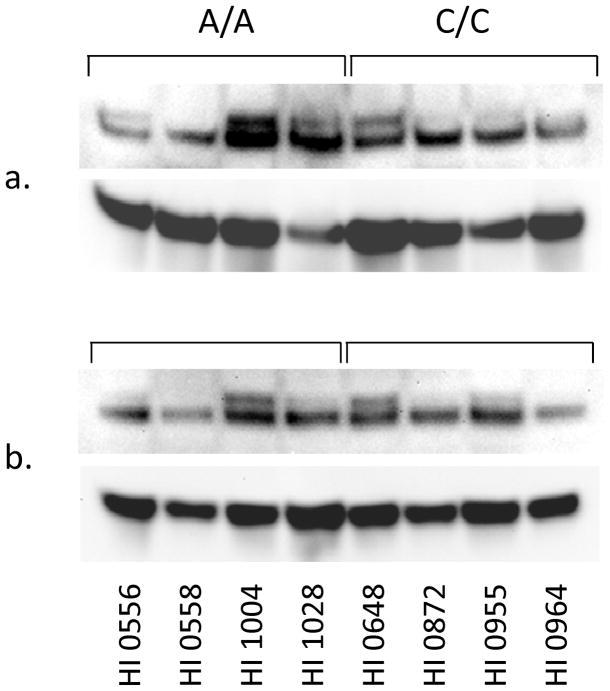
Figure 2.
Western blot analysis for the RAGE in lymphoblastoid cells expressing different isoforms of the Glo1. Equal amounts of proteins (50 μg) from particulate (a) and soluble (b) fractions were resolved on a 10–20% gradient SDS-PAGE followed by transfer onto a nitrocellulose membrane. The nitrocellulose membrane was probed with an anti-RAGE (rabbit polyclonal) antibody, and the blot was also probed with actin as a loading control.

