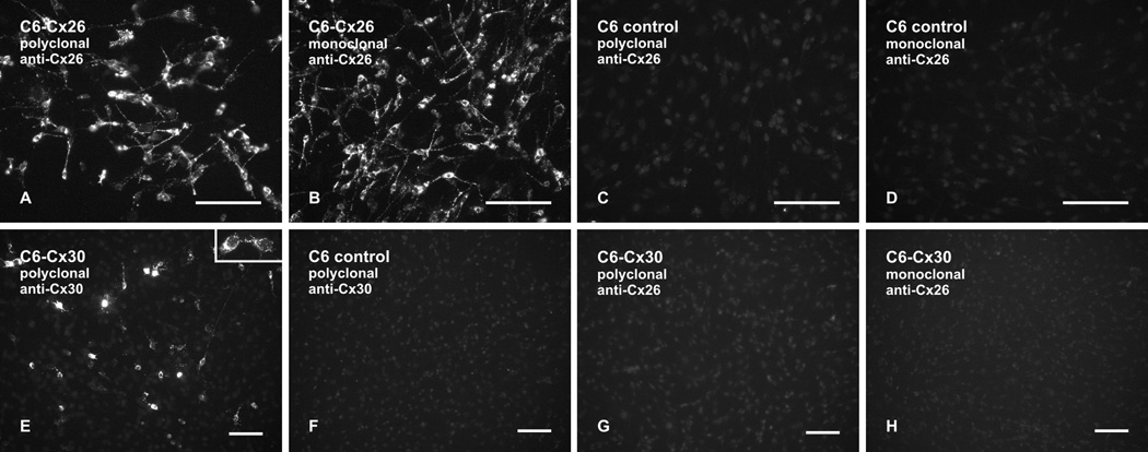
FIG. 2.
Immunofluorescence labelling of Cx26 and Cx30 in C6 glioma cell cultures. (A–D) C6 glioma cells stably expressing Cx26 show immunolabelling with polyclonal anti-Cx26 Ab51-2800 (A) and with monoclonal anti-Cx26 Ab33-5800 (B), and control non-transfected C6 cells show absence of labelling with these antibodies (C,D). (E,F) C6 glioma cells transiently transfected with Cx30 show intracellular labelling (E) and punctate labelling at cell-cell appositions (E, inset) with polyclonal anti-Cx30 Ab71-2200. Control non-transfected C6 cells show an absence of labelling with polyclonal anti-Cx30 Ab71-2200 (F). (G,H) C6 glioma cells transiently transfected with Cx30 show an absence of Cx30 detection with either polyclonal anti-Cx26 Ab51-2800 (G) or monoclonal anti-Cx26 Ab33-5800 (H). Scale bars: 100 µm.

