FIG. 3.
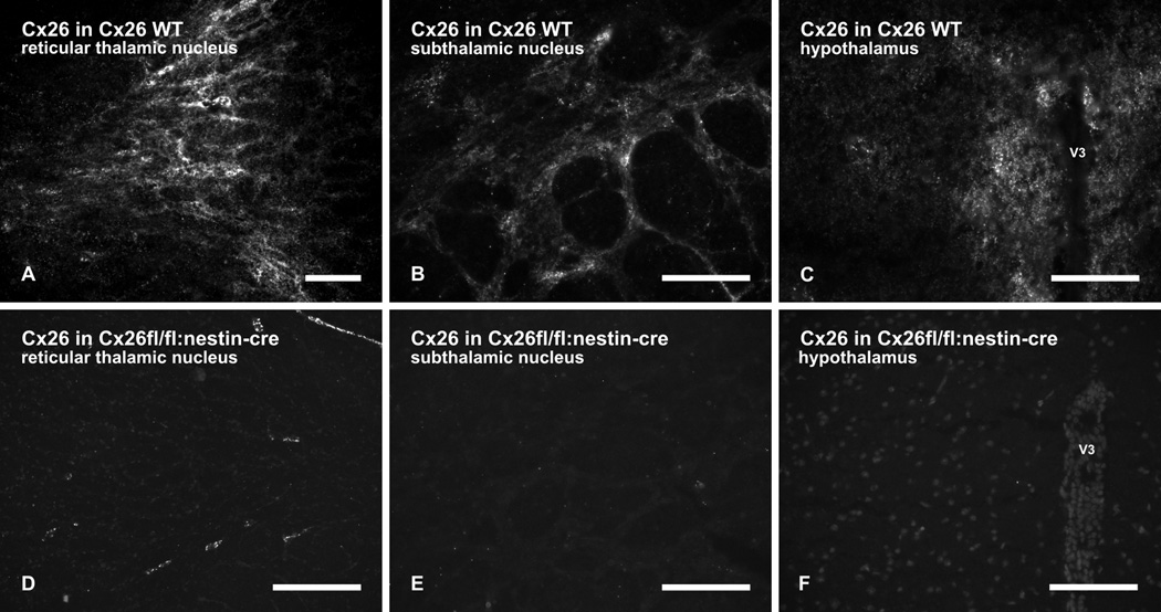
Immunofluorescence labelling of Cx26 in brain regions of wild-type (WT) and Cx26fl/fl:Nestin-Cre adult mice. (A–C) Patches of moderate to dense punctate immunolabelling of Cx26 in wild-type mice is shown in the reticular thalamic nucleus (A), in the subthalamic nucleus (B) and the hypothalamus (C) in the vicinity of the third ventricle (V3). (D–F) Absence of immunolabelling in brain regions of Cx26fl/fl:Nestin-Cre mice is shown in the reticular thalamic nucleus (D), subthalamic nucleus (E) and hypothalamus (F). Some Cx26-puncta in D are associated with leptomeninges (see Fig. 5). Scale bars: A,C,F, 100 µm; B,D, 50 µm; E, 200 µm.

